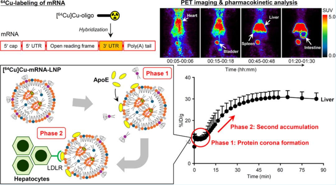
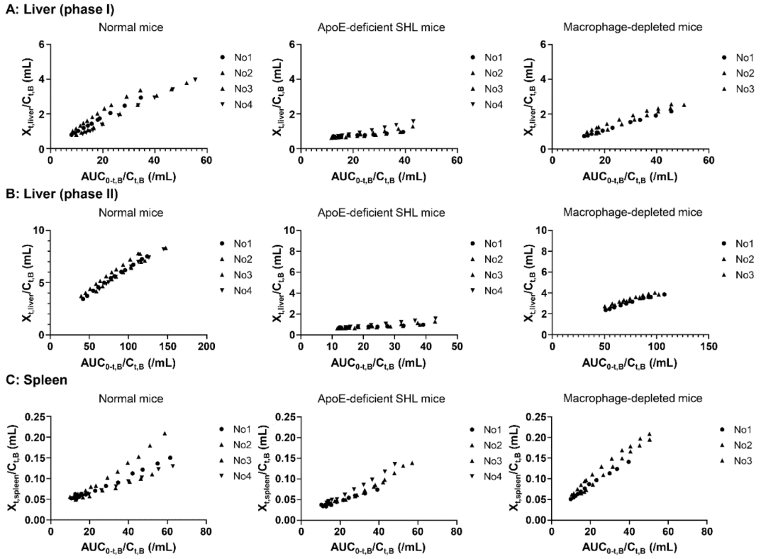
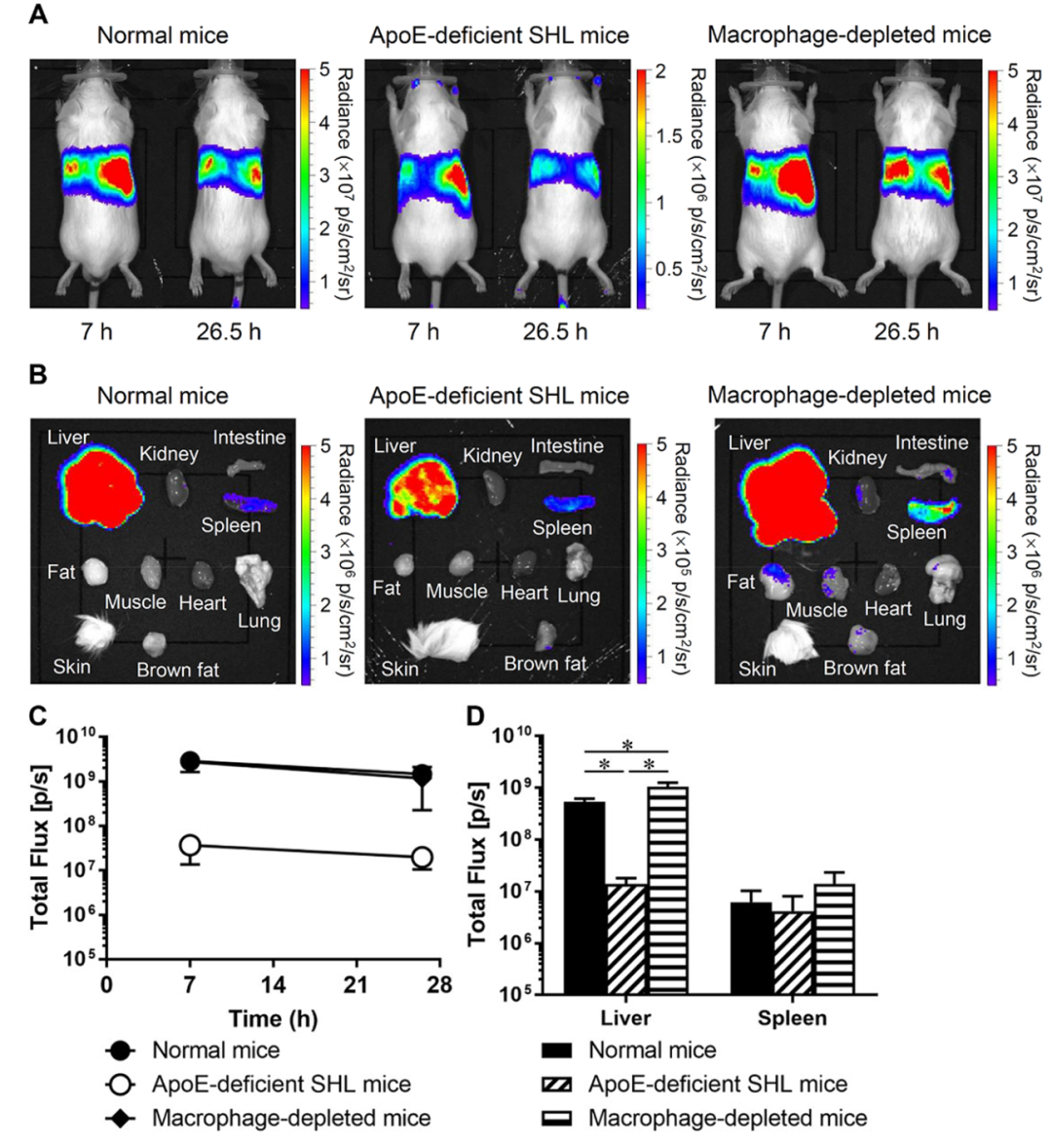
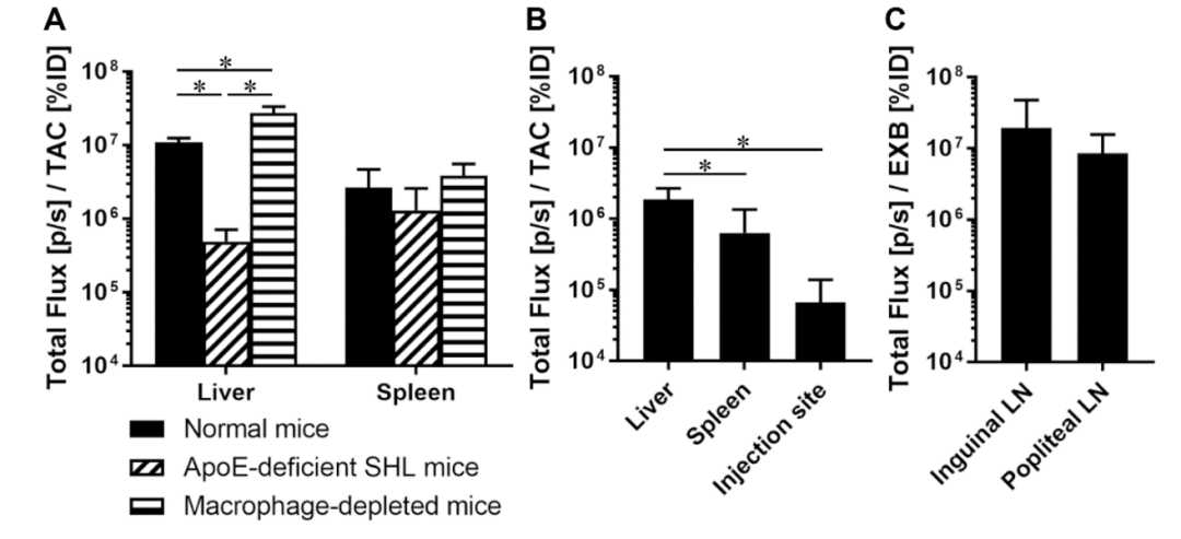
近期,日本长崎大学Hidefumi Mukai团队在ACS Applied Materials & Interfaces发表题为“Positron Emission Tomography-Based Pharmacokinetics of mRNA-Lipid Nanoparticles: A Study Quantifying the ApoE and Macrophage Contribution”的研究。本研究通过开发一种64Cu标记mRNA-LNPs的制备方法,并利用PET技术对其在正常小鼠、ApoE缺陷小鼠和巨噬细胞耗竭小鼠中的药代动力学进行了系统研究,旨在阐明mRNA-LNPs在体内的动态行为及其与蛋白表达的关系。
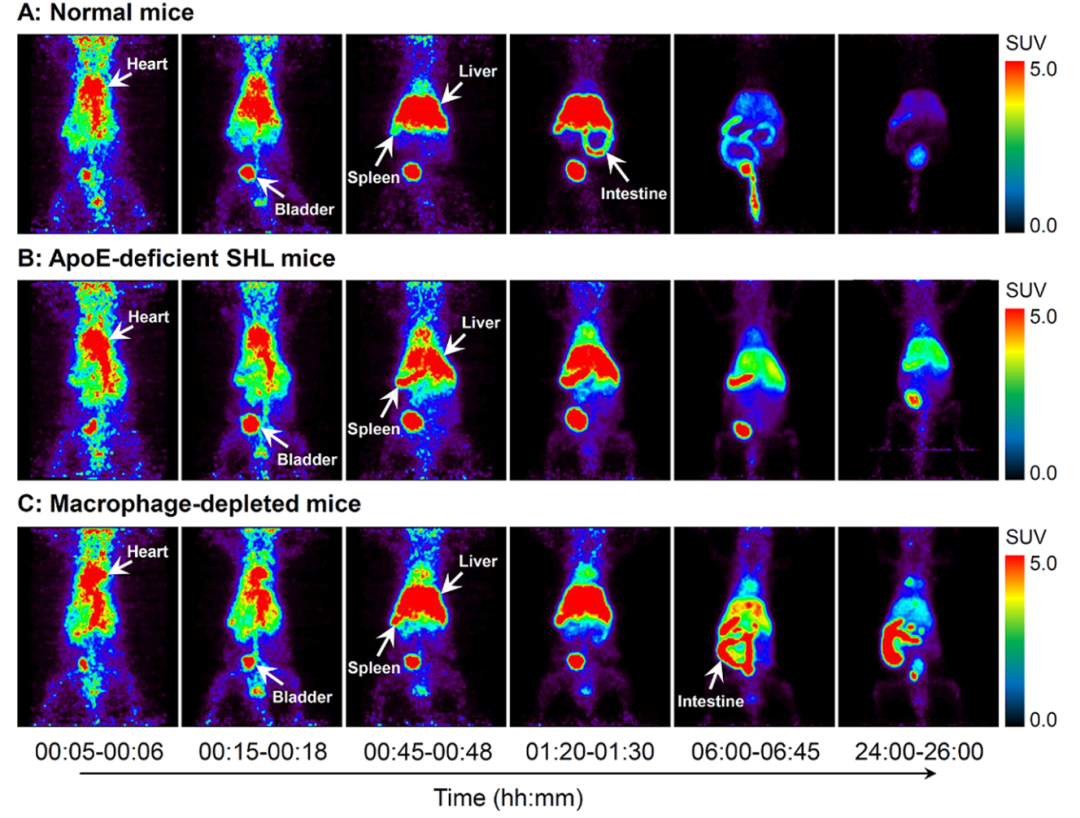
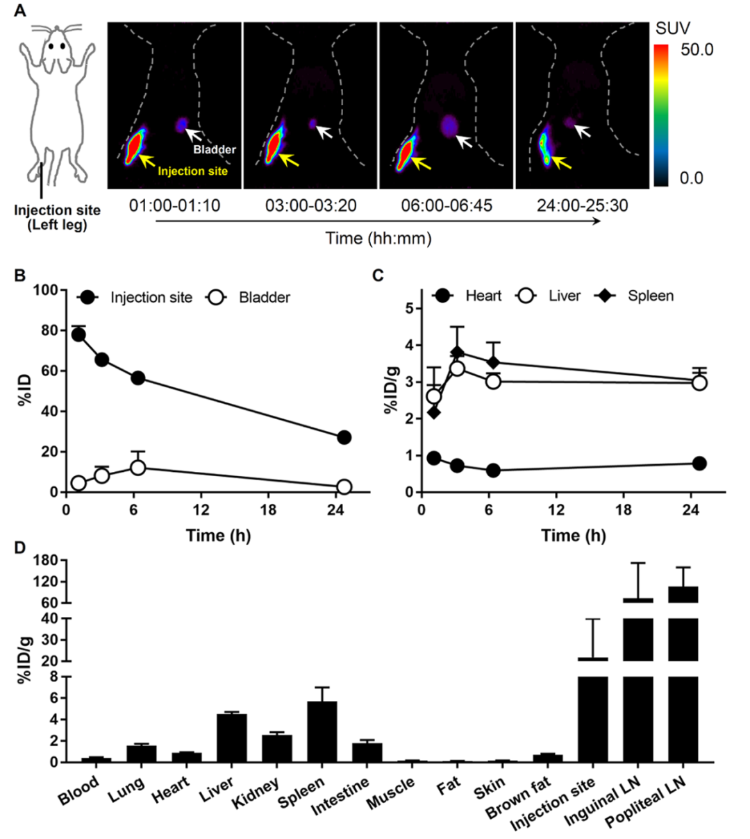
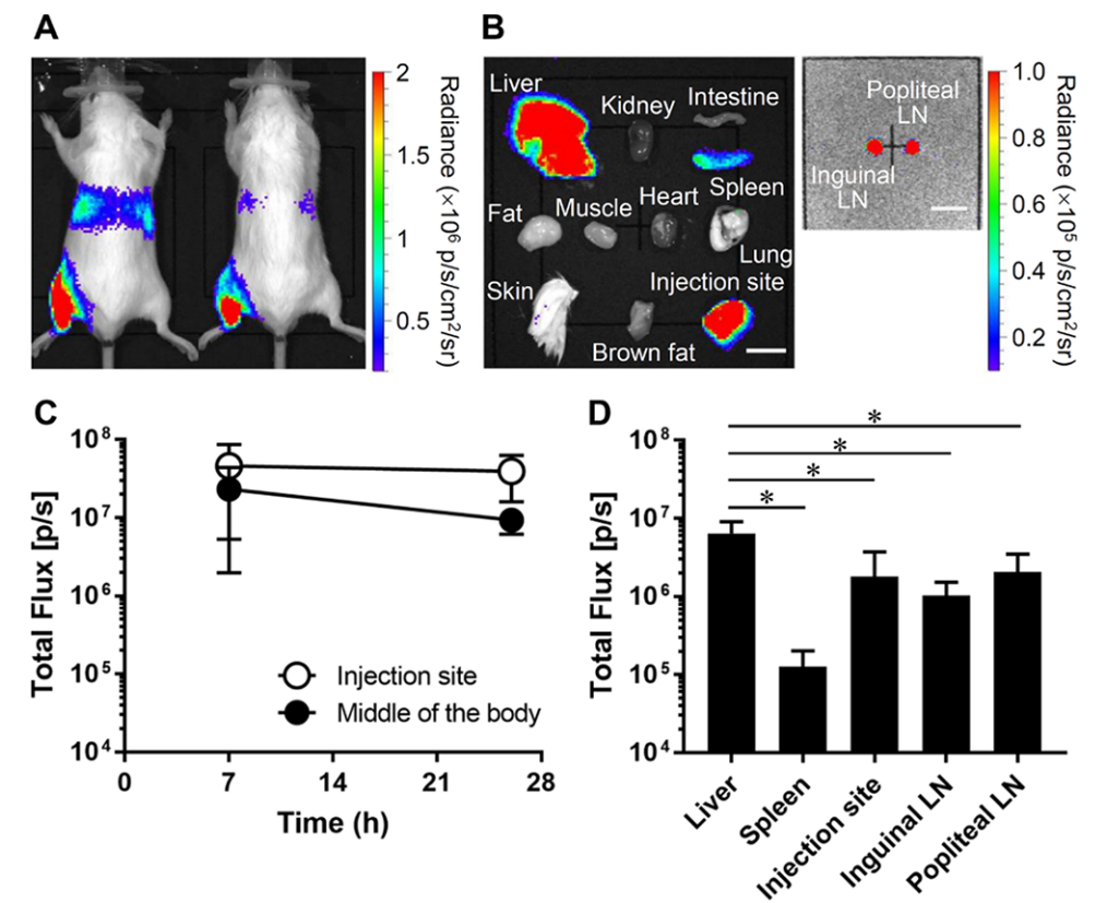
Figure 1. PET images of mice postintravenous injection of [64Cu] Cu-mRNA-LNP. Representative maximum intensity projection PET images of normal mice (A), ApoE-deficient mice (B), and macrophage-depleted mice (C) postinjection of [64Cu] Cu-mRNA-LNP.
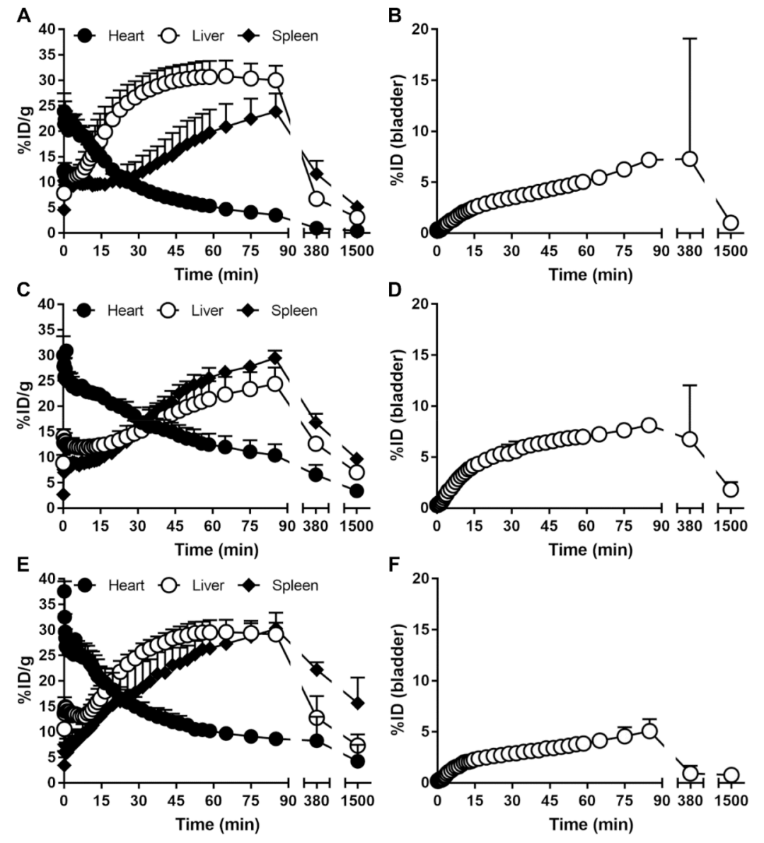
Figure 2. Time-activity curves in tissues of mice postinjection of [64Cu]Cu-mRNA-LNP. The time-activity curves in tissues of normal mice (A and B), ApoE-deficient mice (C and D), and macrophage-depleted mice (E and F) for [64Cu]Cu-mRNA-LNP. Each value represents the mean + SD (n = 3-4).
参考文献:Mohri, Kohta, et al. "Positron Emission Tomography-Based Pharmacokinetics of mRNA–Lipid Nanoparticles: A Study Quantifying the ApoE and Macrophage Contribution." ACS Applied Materials & Interfaces (2025).